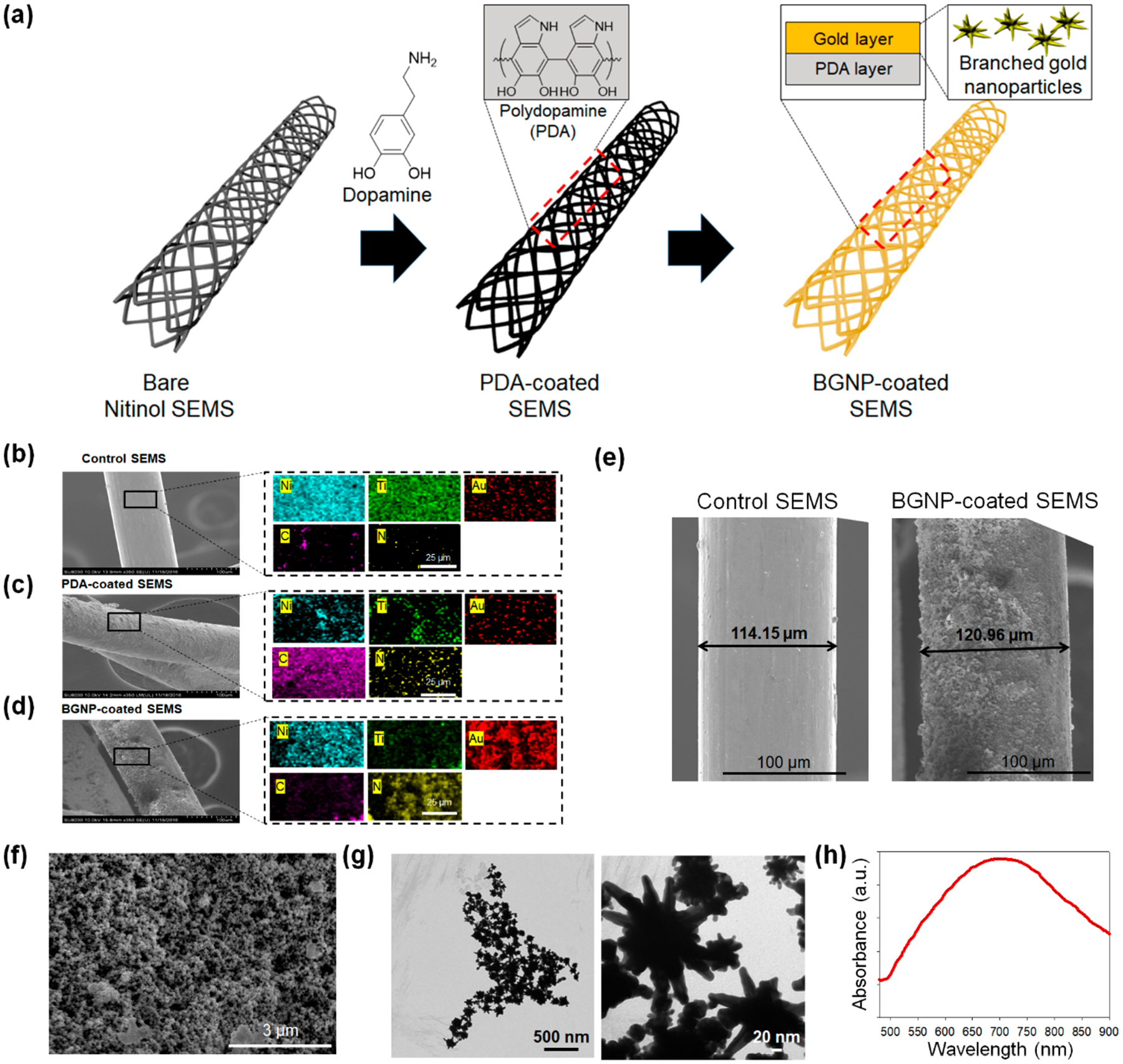
Figure 1.

(a) Schematic illustration of the preparation process for branched gold nanoparticles (BGNP)-coated self-expandable metallic stents (SEMS). The cationic polymer was coated on the surface of SEMS through polydopamine (PDA) coating, and BGNPs were sequentially deposited on the polymer layer. Scanning electron microscopy (SEM) and energy-dispersive X-ray spectroscopy (EDS) mapping analysis of (b) bare SEMS, (c) PDA-coated SEMS, and (d) BGNP-coated SEMS (blue, green, red, pink, and yellow indicate nitinol, titanium, gold, carbon, and nitrogen, respectively). (e) SEM image of bare SEMS and BGNP-coated SEMS. SEM images were analyzed by ImageJ software (U.S. National Institutes of Health, Bethesda, MD) to measure the coating thickness. (f) High-magnification SEM image of the surface of BGNP-coated SEMS. (g) Transmission electron microscopy (TEM) image and (h) UV–vis absorption spectra of BGNP extracted from the surface of BGNP-coated SEMS.
