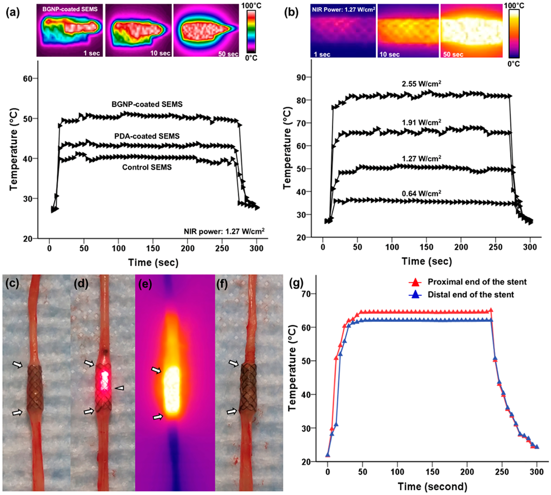
Figure 2.

Infrared thermal camera images and temperature change measurement of (a) bare self-expandable metallic stents (SEMS), polydopamine (PDA)-coated SEMS, and branched gold nanoparticles (BGNP)-coated SEMS under irradiation with an 808 nm laser at 1.27 W/cm2 and (b) BGNP-coated SEMS under irradiation at four different laser powers (0.64, 1.27, 1.91, and 2.55 W/cm2). Photographs and a photothermal image of the rat esophagus with a BGNP-coated stent (arrows) (c) before, (d, e) during, and (f) after NIR laser irradiation using an optical fiber (arrowhead, d). (g) Graph showing temperature changes at the proximal and distal ends of the stent under NIR laser irradiation at 1.91 W/cm2.
