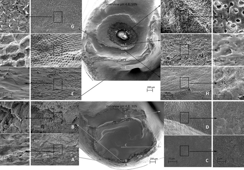
Fig. 3.
SEM pictures of cup-shaped defects from a 50N specimen exposed to pH 4.8 (top, cup-shaped lesion extending into dentine) and a 30N specimen exposed to pH 4.8 (bottom, cup-shaped lesion limited to the enamel). Overview magnification, ×50. The frames indicated A–J show enlargements of areas in the cup-shaped lesion. Magnification, ×2,000 and ×5,000. In the overview cup shape lesions can be observed by multiple cavities and partly overlapping grooves, where on the surface a terrace structure can be observed with indented layering. A, E The enamel on the top of the cusp is slightly polished, the enamel is softened, and the solved components may exhibit a smearing action resulting in less friction and damage. B Enamel prism separated from each other. Separation of prisms will facilitate a deep penetration of acid. F “Polished” key-hole structures can be seen. C, D, G The damaged prisms are eroded/abraded and compressed, and a smear layer seems to arise. H, I In the lowest part of the formed cup-shaped lesion the surface is eroded/abraded and compressed, and a more pronounced smear layer is visible. J Exposed dentine.

