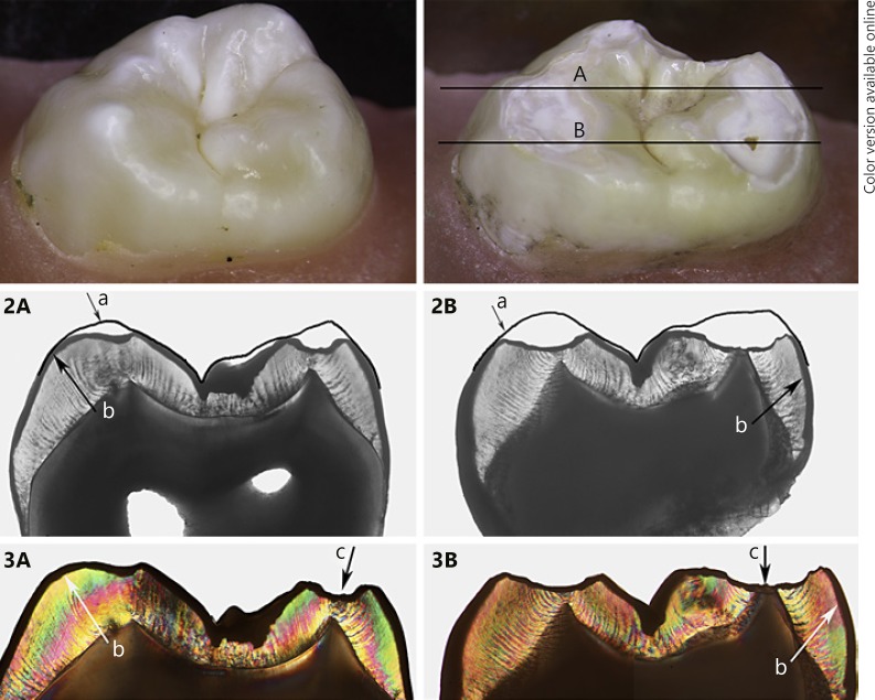
Fig. 4.
Stereomicroscopic images in the top row show a sound specimen (left) and after exposure (right) of a specimen from the pH 4.8/50N group, with lines A and B indicating the location of the 100-µm sections imaged below. Sections assessed by transmitted light microscopy (2A, 2B) and polarized light (3A, 3B): (a) indicates the outline of the original shape of the cusps, (b) the demineralized top surface, and (c) the “deepest” point of the cup-shaped lesion at the location of the dentine “cusp” tip.

