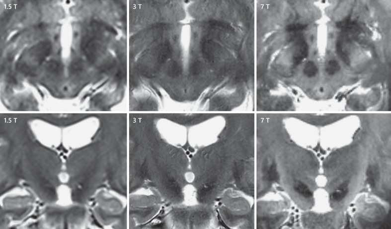
Fig. 1.
Axial and coronal midbrain sections showing the dorsolateral subthalamic nucleus (STN) on 3 different MRI field strengths. Left upper and lower panel, a 1.5-T T2 sequence; middle upper and lower panel, a 3.0-T T2 sequence; and right upper and lower panel, a 7.0-T T2 sequence. All upper panels are axially oriented, and all lower panels are coronally oriented sections. The hypointense STN signal is situated anterolateral to the red nucleus on axially orientated sections - visible as hypointense almond shaped above the substantia nigra on coronally orientated sections. Every increase in field strength results in clearer STN delineation, and the dorsolateral area can best be evaluated on 7.0-T T2 MRI.

