
Fig. 4.
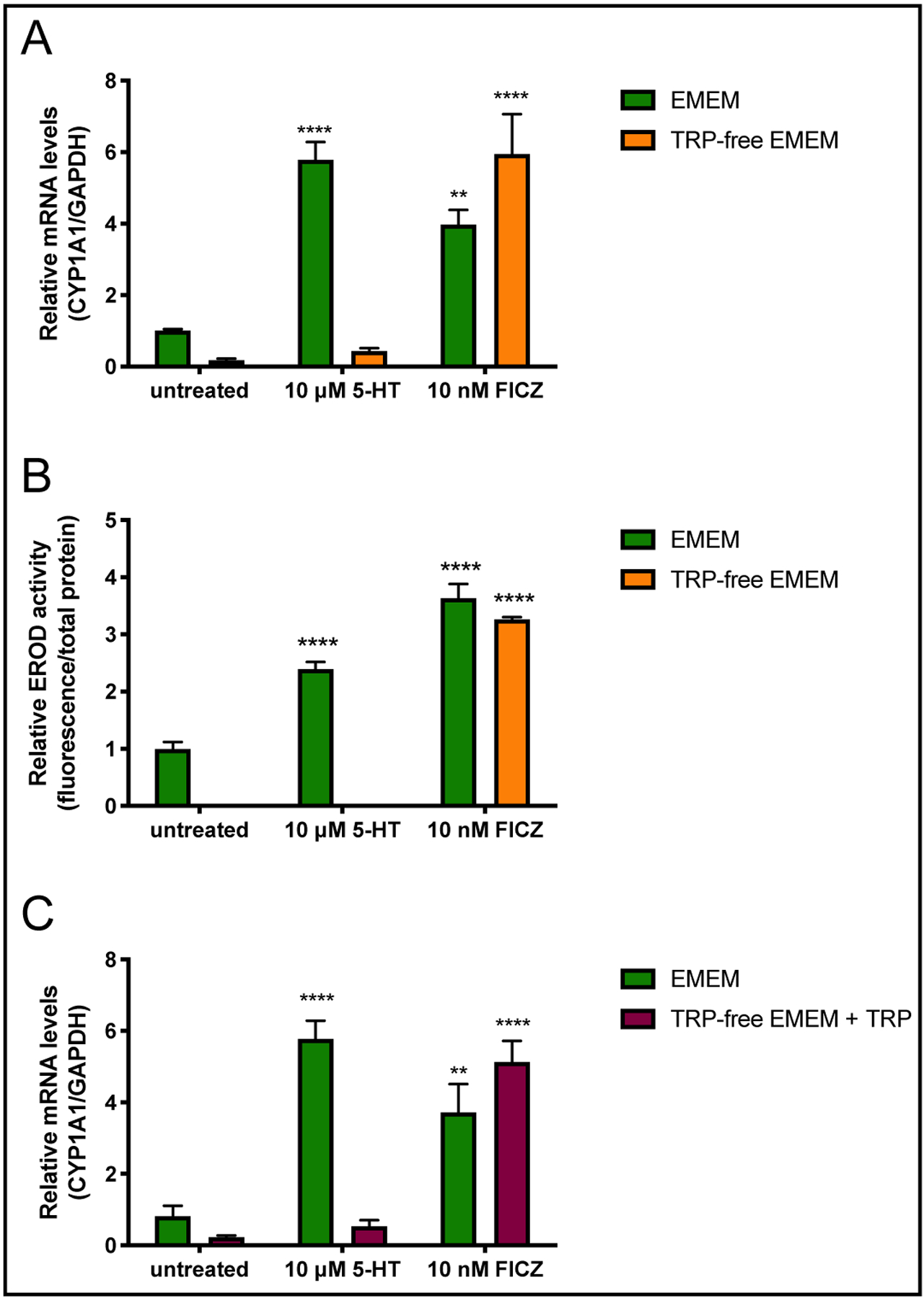
5-HT requires the presence of trace AhR ligands to induce CYP1A1 expression and activity. Caco-2 cells were plated at low density and allowed to differentiate for 10–14 d in medium containing 10% serum before treatments were performed. Cells were treated with 5-HT (10 μM) or FICZ (10 nM) for 8 h in EMEM or TRP-free EMEM. CYP1A1 mRNA (A) was quantified by qPCR and CYP1A1 activity (B) was measured by the ethoxyresorufin-O-deethylase (EROD) assay (n = 3). EROD assays were performed using triplicate wells for each treatment and values were normalized to total protein by Bradford assay. Results are expressed as fold-change mRNA or activity relative to untreated cells in EMEM. (C) The experiment was repeated except recrystallized TRP was added to the TRP-free media immediately prior to treatment. CYP1A1 mRNA was quantified by qPCR and results are expressed as fold-change mRNA relative to untreated cells in EMEM (n = 3). Data analyzed by 2-way ANOVA followed by Dunnett’s multiple comparison’s test. **P<0.01, ****P<0.0001 vs. untreated cells in the same media.