Figure 6.
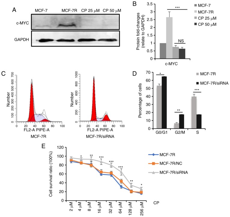
c-MYC affects the sensitivity of cisplatin through regulating the cell cycle. (A) MCF-7R cells were treated with 0, 25 or 50 µM cisplatin for 24 h. After removing the dead cells, cells were lysed. c-MYC expression levels in the indicated cells were determined by western blot analysis. (B) Densitometric analysis of c-MYC in the western blots shown in (A). (C) c-MYC expression was knocked down in MCF-7R cells and flow cytometric analysis was performed to evaluate proportion of cells in the S-phase. (D) Bar chart representing the percentage of MCF-7R cells in the G0/G1, G2/M or S phase, prior to and following c-MYC/siRNA transfection. (E) Cell-viability of MCF-7R with c-MYC knocked down compared with MCF-7R and MCF-7R/NC cells, after treatment of cells with different doses of cisplatin for 24 h (*P<0.05, **P<0.01, ***P<0.001, compared to NC only). Data are presented as the means ± standard deviation of the mean of 3 repeats. *P<0.05, **P<0.01, ***P<0.001. TAM, tamoxifen; siRNA, small interfering; NC, negative control.

