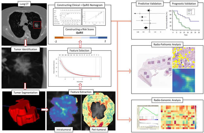
Figure 2:
Overall workflow and pipeline of the project. The first step involved identifying and annotating the primary nodule on the CT scan. Intratumoral and peritumoral textural features were extracted using MATLAB 2016. For the peritumoral region, features were extracted from 0–15mm region outside the tumor and divided into five 3mm peritumoral rings. Feature statistics including mean, median, standard deviation, skewness, kurtosis and range were calculated for each of the individual annular rings. Top features were selected using LASSO feature selection method and used for constructing QuRiS. QuRNom was constructed using prognostic clinical features and QuRiS. QuRiS and QuRNom were validated for prognostic performance and predicting added benefit of adjuvant-chemotherapy. Associations between QuRiS features and spatial patterns of TILs on whole-slide tissue scans were also evaluated, as were associations with mRNA data and underlying immune specific biological pathways.

