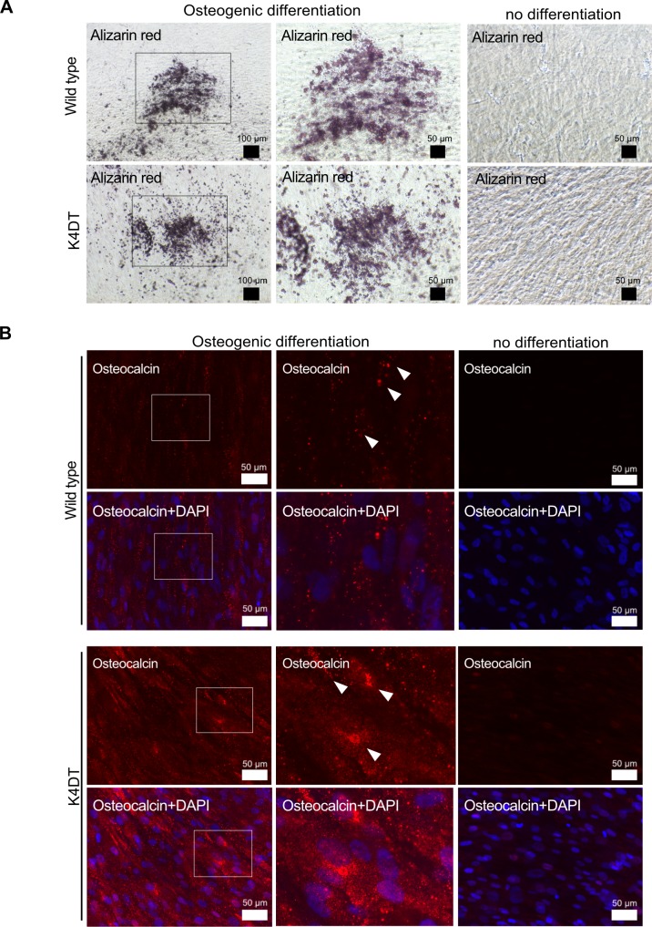
Fig 8. Osteogenic differentiation properties of wild type and K4DT cells.
Representative Alizarin Red S staining (A) and immunostaining (B) with anti-osteocalcin antibody (red) and DAPI staining (blue) of wild type and K4DT cells. For the Alizarin Red S staining, cells were cultured in osteoinduction medium for 17 days; 25 days for the osteocalcin staining. Middle panels show high magnification of the boxed regions in the left panels. White arrows indicate the positive expression of osteocalcin.

