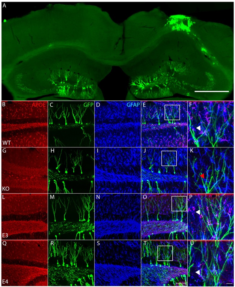
Fig 1. ApoE-expressing astrocytes physically interact with injury-induced GFP-expressing dendrites.
(A) Representative section of the contralateral and ipsilateral cortex and hippocampus 4 weeks after Controlled Cortical Impact and stereotaxic injection of GFP-expressing retroviruses in the dentate gyrus of a wildtype mouse reveals the typical morphology of the injured hippocampus along with efficient long-term labelling of neural progenitor cells. Scale bar 500μm. (B-U) Representative pictures of ApoE (first column, antigen is recombinant human ApoE and can react with human e2, e3 and e4, as well as non-human primate and mouse ApoE), GFP (second column) and GFAP (third column) immunostaining in the dentate gyrus of WT (B-F), ApoE KO (G-K), ApoE3 (L-P) and ApoE4 mice (Q-U). (E, J, O, T) Merged pictures (scale bar 100μm), white squares indicate enlarged area shown in last column (F, K, P, U), white arrowheads designate ApoE-expressing GFAP-positive astrocytes wrapping around GFP-expressing dendrites, red arrowhead designate ApoE-deficient GFAP-positive astrocyte, scale bar 10μm.

