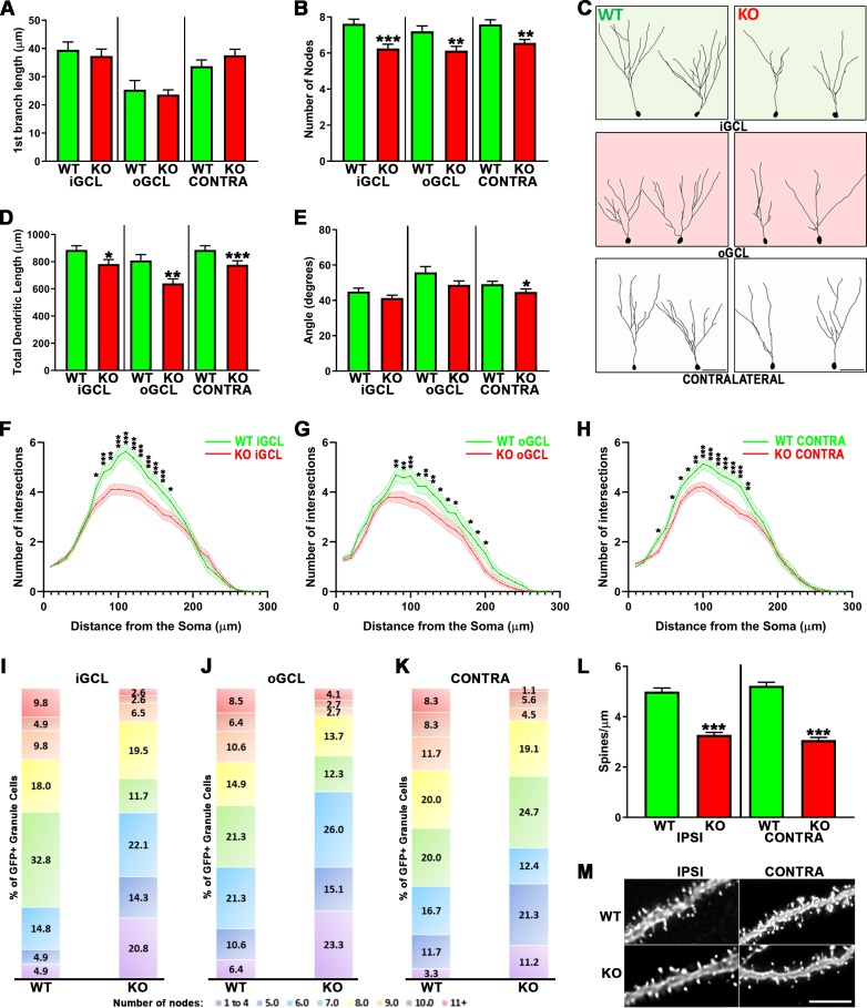
Fig 2. ApoE deficiency leads to impaired dendritic development of injury-induced adult-born granule neurons.
(A) ApoE deficiency did not affect the distance to the first dendritic branching but did significantly affect dendritic complexity (B) in the iGCL (p < 0.001), oGCL (p < 0.01) and contralateral (p < 0.01) dentate gyrus when compared to WT. (C) Representative tracings of GFP-expressing cells found in the iGCL, oGCL and contralateral dentate gyrus from WT and ApoE-deficient injured dentate gyrus. (D) The cumulative dendritic length is significantly attenuated in mouse lacking ApoE when compared to matching Wild Type cells (iGCL: p < 0.05, oGCL: p < 0.01, Contra: p < 0.001). (E) Injury-induced cells found in the contralateral dentate gyrus in the absence of ApoE also showed decreased dendritic span when compared to WT (p < 0.05). Sholl analysis of dendritic arborizations of CCI-induced adult-born granule neurons exposed differences in the dendritic branching number observed in both proximal and distal regions when comparing WT with ApoE-deficient GFP-expressing cells found in the iGCL (F), in the oGCL (G) and in the contralateral dentate gyrus (H). (I-K) In each condition, all traced neurons have been categorized depending on their complexity (≤4…≥11 nodes), highlighting higher proportions of less complex neurons (<4 nodes) in ApoE-deficient, particularly in the oGCL. 4 mice/condition and at least 10 neurons/mouse were analyzed; WT iGCL: 61 cells; WT oGCL: 47 cells; WT Contra: 60 cells; ApoE KO iGCL: 77 cells; ApoE KO oGCL: 73 cells; ApoE KO Contra: 89 cells. Contra = Contralateral dentate gyrus; iGCL = Inner Granule Cell Layer, oGCL = Outer Granule Cell Layer, in the ipsilateral dentate gyrus; WT = Wild Type, KO = ApoE Knock-out. ***p < 0.001. (L) ApoE deficiency leads to significantly reduced spine density in injury-induced adult-born granule neurons when compared to WT cells from both the Ipsilateral and Contralateral sides (p < 0.001). High power representative pictures of dendritic fragments from mature adult-born granule neurons in wildtype and ApoE-deficient (M). 4 mice/condition; Number of dendritic fragments analyzed: WT Ipsilateral = 60, WT Contralateral = 62, ApoE KO Ipsilateral = 87, ApoE KO Contralateral = 53. Scale bar = 5μm.

