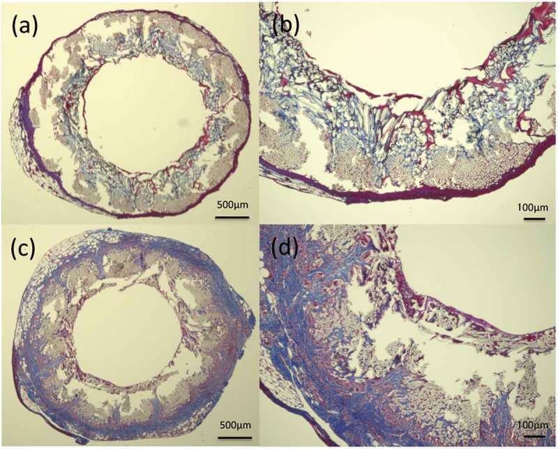
Figure 6.

Histological cross-section images of MTC staining 2 weeks after implantation: (a) gelatin-coated graft and (b) higher magnification of the gelatin-coated graft, (c) SF(Glyc)-coated graft and (d) higher magnification of the SF(Glyc)-coated graft. In the gelatin-coated graft, the presence of collagen fibers was hardly confirmed on the outer periphery and inside the graft. In the SF(Glyc)-coated graft, collagen fibers were mainly gathered around the graft. Moreover, it was observed that they penetrated from the outside to the inside of the graft, also entering through the gaps between the polyester fibers.
