
Figure 3a:
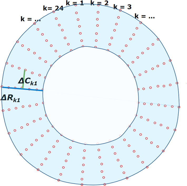
Measurements of radial and circumferential inter-landmark distances at (a) frame 1 (end diastole, assumed as the reference frame) and (b) frame t. Both images depict the seven circumferential rings of landmarks. Subendocardial, midwall, and subepicardial circumferential strain was calculated from the second, fourth, and sixth rings from the center, respectively. ΔRkt shows the distance for radial line k in frame t, while ΔCkt shows the distance for circumferential line k in frame t.