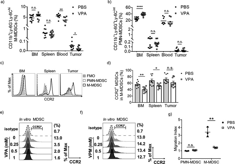
Figure 4.

VPA reduces the infiltration of M-MDSCs in EL4 tumors.
Flow cytometric analyses of BM, spleen, blood, and tumors from PBS- or VPA-treated EL4 tumor-bearing mice 14 days after EL4 inoculation. The proportion of each population is plotted; (a) M-MDSCs (CD11b+Ly-6G−Ly-6Chi) and (b) PMN-MDSCs (CD11b+Ly-6G+Ly-6Cint). Data represent the means of two (blood) or three (BM, spleen, and tumor) independent experiments (*p < .05, **p < .01, and ****p < .0001 by Student’s t test). (c) CCR2 expression levels in PMN-MDSCs and M-MDSCs from PBS-treated tumor-bearing mice assessed by flow cytometry. (d) The proportion of CCR2+ MDSCs among total M-MDSCs in BM, spleen, and tumor from PBS- or VPA-treated mice. Data represent means ± S.E.M. of three independent experiments (*p < .05, **p < .01 by Student’s t test). (e, f) CCR2 expression on in vitro MDSCs following treatment with different concentrations of VPA or VPM. Data are one experiment representative of two independent experiments. (g) BM cells were harvested from EL4 tumor-bearing mice treated with PBS or VPA. The migration index was calculated as a ratio of the number of migrated cells treated with chemokines CCL2/CCL7 to that in non-treated wells. Data represent means ± S.D. in triplicate of one representative experiment from two independent experiments. (**p < .01 by Student’s t test)
