
Figure 8.
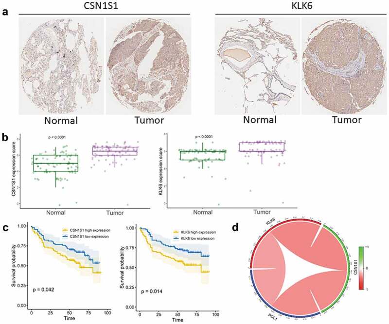
Validation of the gene signature in clinical tissue samples (a) Representative images of IHC staining for CSN1S1 and KLK6 expression in LUSC tissues and adjacent non-tumor tissues. (b) Protein expression scores in LUSC tissues and normal lung tissues. (c) Kaplan–Meier analysis of overall survival according to CSN1S1 and KLK6 expression levels. (d) Correlation of PD-L1 expression with KLK6 and CSN1S1 expression.