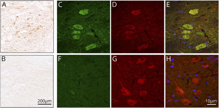
Figure 2. Detection of KLHL11-ab by immunohistochemistry on rat tissue.
Immunohistochemistry on rat brain sections showing cytoplasmic staining of neurons of deep cerebellar nuclei incubated by a representative patient's serum with KLHL11-ab (A) and a negative control (B). Scale bar = 200 μm. Panel C shows the reactivity (green) of a representative patient's serum with HEK293 cells expressing KLHL11. No immunoreactivity is observed with a serum control (F). Panels D and G show the reactivity (red) of a commercial KLHL11-ab. Panels E and H show the merged reactivities of the indicated samples (patient's and control samples) with the commercial KLHL11-ab, showing a perfect colocalization with patient's antibodies (E). Scale bar = 10 μm.

