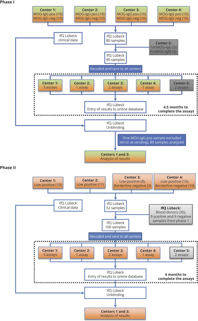
Figure 1. Flowchart showing phases I and II of this study.
Center 1 (Innsbruck) performed 5 assays (live CBA-IF MOG-IgG (H + L), live CBA-IF MOG-IgG(Fc), live CBA-FACS MOG-IgG(Fc), live CBA-IF MOG-IgM, and ELISA MOG-IgG); center 2 (Mayo Clinic) performed 1 assay (live CBA-FACS MOG-IgG1); center 3 (Oxford) performed 2 assays (live CBA-IF MOG-IgG (H + L) and live CBA-IF MOG-IgG1); center 4 (Sydney) performed 1 assay (live CBA-FACS MOG-IgG (H + L)), which was repeated twice; center 5 (Euroimmun) performed 2 assays (fixed CBA-IF MOG-IgG(Fc) and ELISA MOG-IgG(Fc)). CBA = cell-based assay; FACS = fluorescence-activated cell sorting; IF = immunofluorescence; IfQ = Institute for Quality Assurance; Ig = immunoglobulin; MOG = myelin oligodendrocyte glycoprotein.

