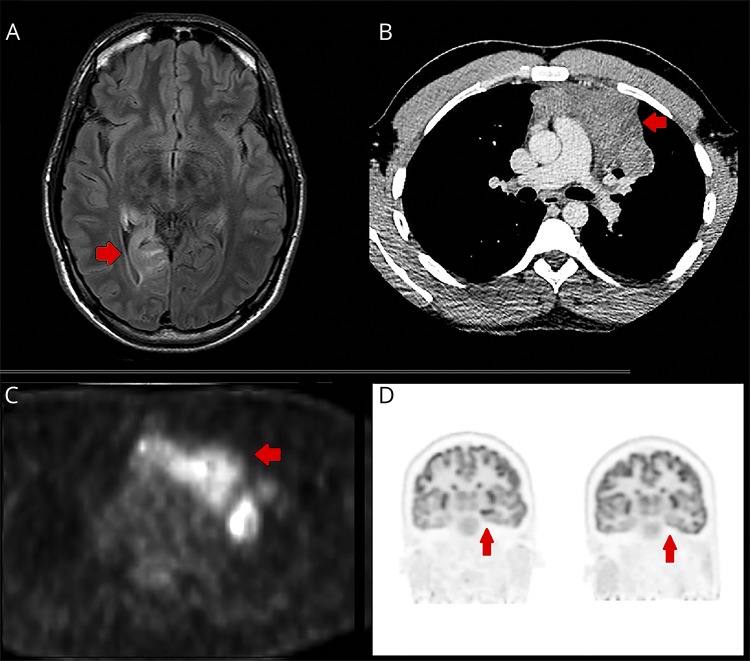
Figure. Imaging investigations.
(A) Axial T2 fluid attenuated inversion recovery (FLAIR) MRI of the brain showing hyperintensity in the right medial temporal lobe at the age of 22 years. (B) Large anterior mediastinal mass diagnosed at the age of 28 years as demonstrated on CT chest. (C) Large anterior mediastinal mass (age 28) demonstrating high glucose avidity on PET-CT. (D) Left: Markedly increased metabolism in the left anteromesial temporal lobe and hippocampus. Right: Resolution of those changes 11 months after resection of the mediastinal tumor.

