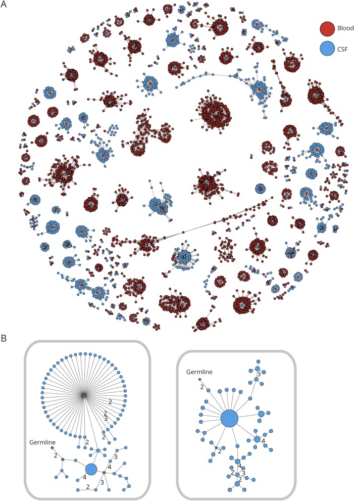
Figure 2. Intrathecal somatic hypermutation in patients with leucine-rich, glioma-inactivated 1 (LGI1) antibody encephalitis.
(A) Data from 1 representative patient of 6 LGI1 patients showing all clusters of clonally related immunoglobulin heavy chain variable region, which are shared between peripheral blood (PB) and CSF B cells. Each red dot represents ≥1 identical PB sequence, and each blue dot ≥1 identical CSF sequence. Two dots connected by a line differ from each other by a Hamming distance of 1 in their CDR3 region on the nucleotide sequence level. Clusters of related sequences are grouped together. (B) Two Ig lineage trees of CSF B cells from 1 patient are shown. Each dot represents 1 sequence, and its size correlates with the number of times this sequence could be found. Two dots connected by a line differ from each other by 1 nucleotide in the CDR3 region unless marked otherwise. Putative germline nodes are labeled; lineage intermediates not found in the sequencing data were calculated and are labeled in gray.

