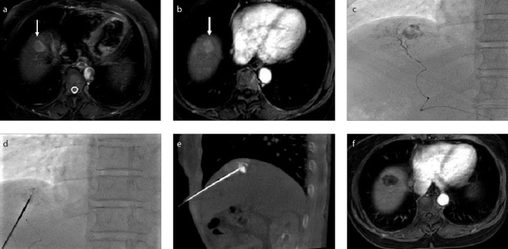
Figure 1. a–f.
A hepatocellular carcinoma (HCC) lesion near the diaphragm. Contrast-enhanced magnetic resonance images (a, b) demonstrate the lesion (arrow). Transarterial chemoembolization (TACE) (c) was performed to mark the lesion. Image (d) shows needle puncture performed under fluoroscopy. Cone beam CT (e) confirmed the position of the needle within the lesion. Contrast-enhanced MRI (f) confirmed complete necrosis of the lesion after radiofrequency ablation (RFA).

