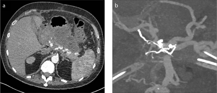
Figure 3. a, b.
CT image (a) demonstrating pseudoaneurysm arising from the proximal common hepatic artery. CT reconstruction (b) demonstrating embolization performed from a small celiac collateral with closure of both afferent and efferent supply to the pseudoaneurysm, whilst preserving the main right hepatic artery.

