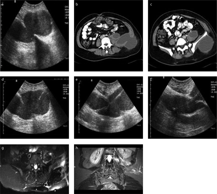
Figure 1. a–h.
Diagnostic, procedural and follow-up imaging of a percutaneously treated tuberculous psoas abscess. Preprocedural US (a) and contrastenhanced venous phase transverse abdominal CT (b, c) images reveal a multiloculated collection involving left psoas muscle extending to the left flank region, neighbouring left iliac bone. US images (d–f) captured during procedure show introduced 18 G Seldinger needle (d) and 0.035-inch guidewire (e), followed by insertion of 12 F catheter (f) into the abscess cavity. Follow-up T2-weighted fat saturated transverse and coronal MRI images (g, h) demonstrate minimally increased signal intensity in the left psoas muscle and soft tissues surrounding the left iliac bone with no residual or recurrent cavity.

