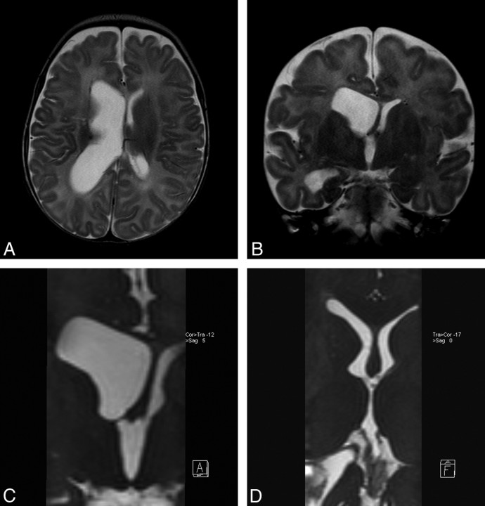
Fig 5.
Case 4. A, Axial TSE T2 image through the lateral ventricle demonstrates unilateral right lateral ventriculomegaly. B, Coronal TSE T2 image through the foramen of Monro falsely demonstrates a free communication between the right lateral ventricle and the third ventricle. C, Coronal reformatted sagittal 3D-CISS image points out a complete membranous obstruction in the right foramen of Monro. D, Follow-up axial-oblique reformatted image revealing complete removal of obstructing membrane and normal-appearing lateral ventricles.

