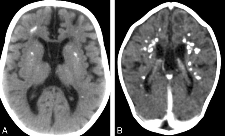
Fig 2.
Cerebral calcifications. A, Axial nonenhanced CT image of case 1 shows numerous punctuate calcifications within the basal ganglia and the cerebral white matter, a pattern typical in patients with AGS. B, Contrast-enhanced CT scan (case 19) shows large calcifications in the white matter. Although the CT examination was performed in the acute phase of the disease, no signs of contrast enhancement are seen. Atrophy, microcephaly, and areas of hypoattenuation in the periventricular white matter are also evident.

