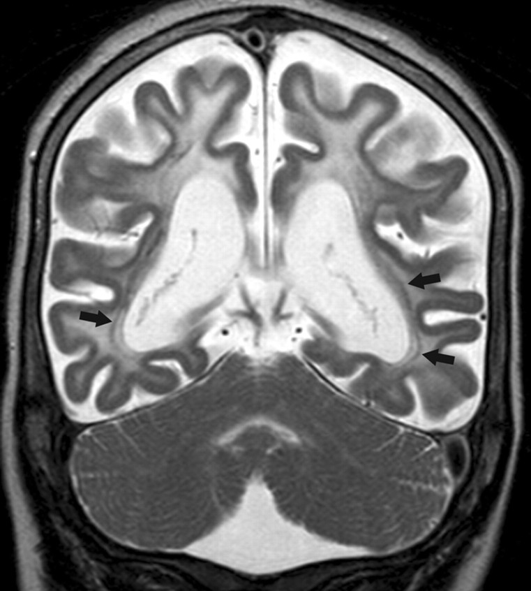
Fig 3.
Coronal fast spin-echo T2-weighted MR image (1.5T, TR = 5022 ms, TE = 100 ms) of case 10 shows a diffuse signal-intensity abnormality of the cerebral lobar white matter. Note that the cerebellar white matter and the optic radiations (arrows) are spared, whereas the subcortical U-fibers are involved. The cortex shows an even thickness. Severe cerebral atrophy is evident.

