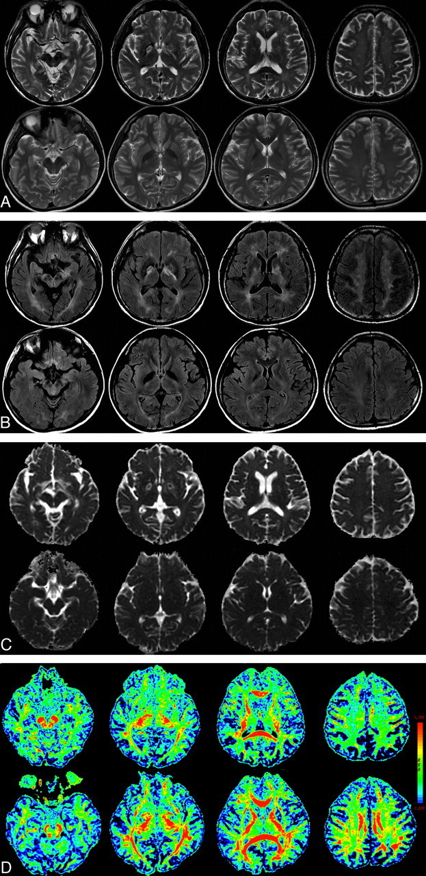
Fig 2.

A 57-year-old man with delayed neuropsychiatric syndrome after CO intoxication (upper) and a healthy control (lower). A and B, Axial T2-weighted images (A) and FLAIR MR images (B) show symmetric necrosis of the bilateral globus pallidus and extensive hyperintensity of the subcortical WM. C, ADC map. D, The FA map of DTI reveals clearly reduced fiber integrity of the corresponding WM. The color bar represents the FA values.
