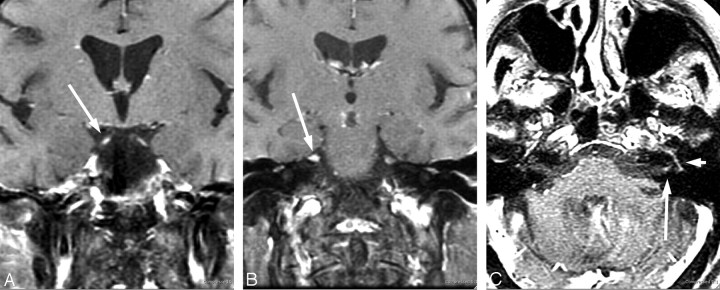
Fig 5.
Evolving cranial neuritis. A 71-year-old woman with headache, malaise, fever, and diplopia. Initial coronal postcontrast T1 MR imaging (A and B) with enhancing bilateral third and fifth cranial nerves. C, Nine days later, she developed left facial palsy with enhancing fundal tuft and labyrinthine and tympanic segments of the seventh cranial nerve. The patient had CSF pleocytosis with positive Lyme-EIA and Western blot in both serum and CSF and CSF Lyme PCR-negative findings. Resolution occurred with intravenous ceftriaxone therapy.

