
Fig 4.
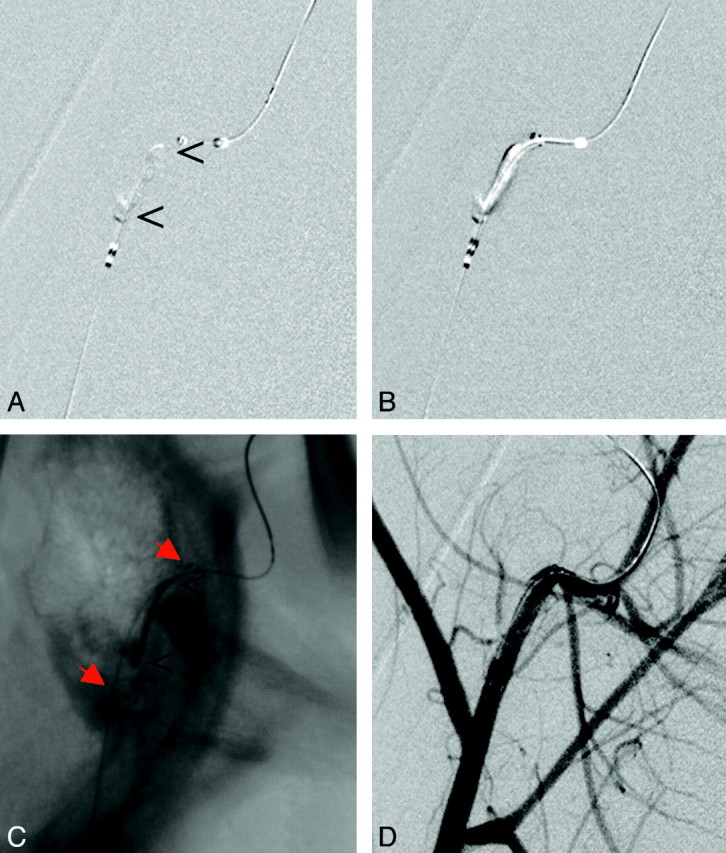
A, Occlusion of the maxillary artery in proximity to a sharp bend of the vessel. B and C, The stent (red arrows) is placed across the thrombus (B) and deployed (C). Again, the thrombus (open arrowheads) is compressed to the side contralateral to the initial passing procedure and is stabilized there (C). D, Follow-up angiogram reveals nearly complete recanalization.