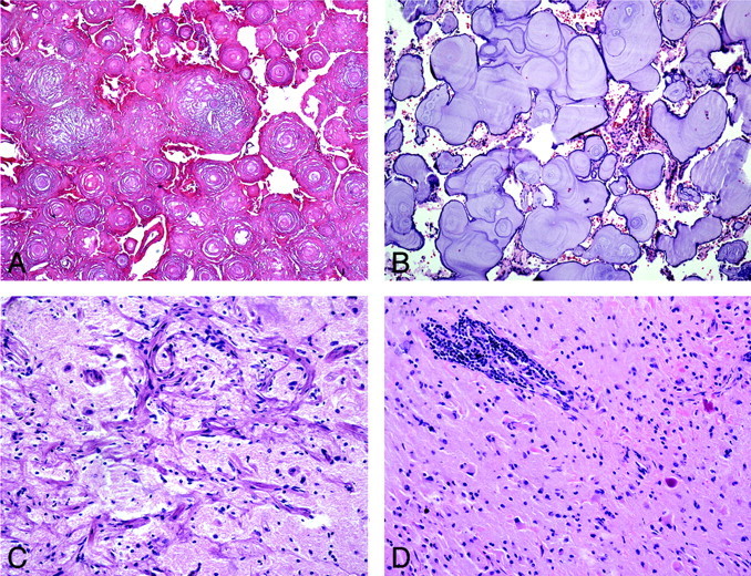
Fig 4.

Atypical and unusual features of CAPNON. A, Areas of coalescent concentric lamellar calcifications without intervening chondromyxoid matrix or cells (H&E, original magnification ×100). B, More basophilic amorphous lamellar calcifications without intervening chondromyxoid matrix and with rare meningothelial cells (H&E, original magnification ×100). C, Adjacent cortical region showing meningioangiomatosis (H&E, original magnification ×200). D, Surrounding parenchyma with prominent perivascular lymphocytic infiltrates and Rosenthal fibers (H&E, original magnification ×200).
