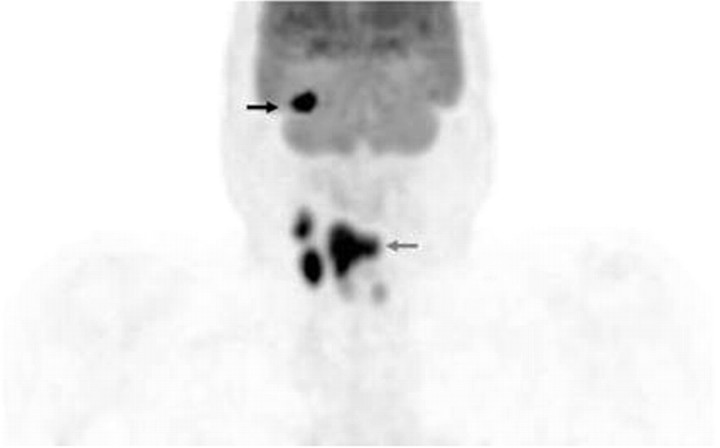
Fig 1.
Volumetric FDG-PET image demonstrates increased uptake in the supraglottic/hypopharyngeal region consistent with the primary tumor (gray arrow). Additionally noted are multiple foci of FDG uptake in right-sided cervical lymph nodes, consistent with metastatic disease. There is an additional focus of intense uptake in the region of the maxilla, suggestive of metastatic disease (black arrow; Fig 2). Faint and symmetric uptake in the midline is consistent with physiologic uptake in the vocal cords.

