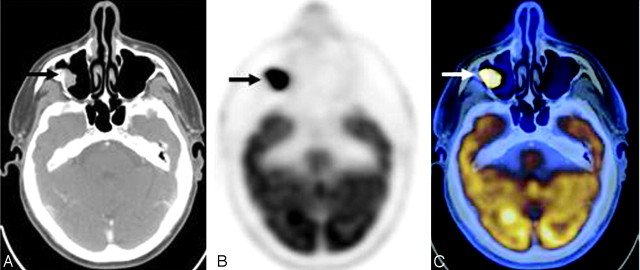
Fig 2.
Fusion (A), PET (B), and axial CT (C) images demonstrate a focus of intensely increased FDG uptake (arrow) corresponding to a soft-tissue attenuation projecting off the epithelial surface of the right maxillary sinus, highly suggestive of a malignant process. However, histopathologic examination revealed this mass to be a schneiderian papilloma.

