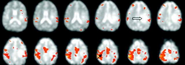
Fig 3.
Patient 12 with a metastatic tumor. Images show identification of the motor cortex (heavy solid arrow) during bilateral finger tapping. The motor area is identified from the characteristic signal peaks. Selection of a voxel in the motor area (solid arrow) highlights both motor areas and the supplementary motor area but does not highlight voxels in the tumor mass.

