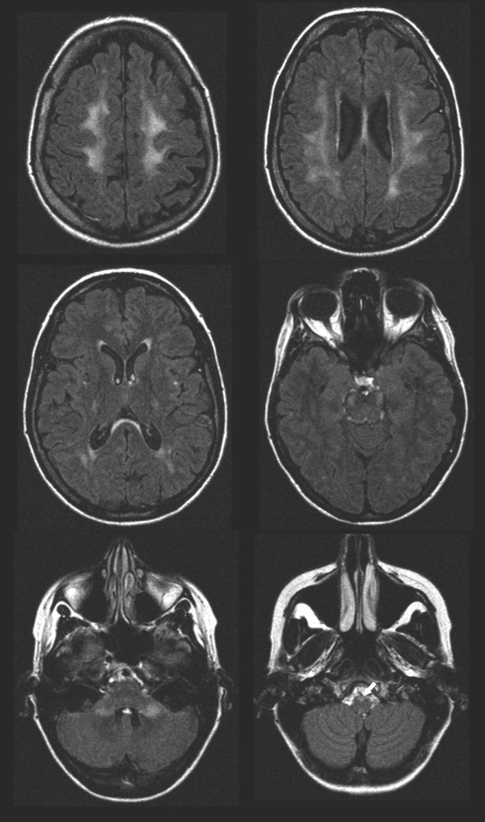
Fig 5.

Fluid-attenuated inversion recovery images of the brain in the same patient as in Fig 2. Increased white matter SI can be seen beneath the motor cortex and in the upper part of the parietal lobes. The SI changes can be followed along the corticospinal tract including the pyramids (arrow) and in the cerebellar peduncles. A periventricular rim with less increased signal intensity is characteristic of adult-onset ADLD with autonomic symptoms.
