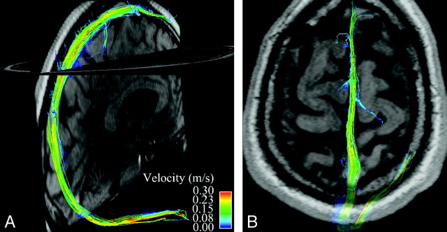
Fig 1.
Whole-brain venous imaging in a healthy volunteer. A maps streamlines of venous blood flow in the superior sagittal and right transverse sinuses onto a midline sagittal magnitude image. Streamlines are imaginary lines aligned with local vector fields and represent the flow field at a given moment in the cardiac cycle. They are color coded for velocity. B demonstrates flow in the superior sagittal sinus at the vertex with a semitransparent axial magnitude image provided for orientation.

