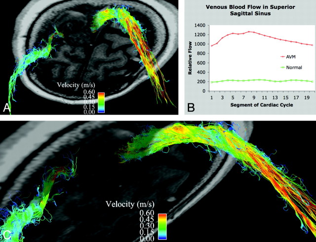
Fig 3.
Venous drainage in a patient with an AVM. A is a slightly oblique view of an axial section through the left frontoparietal AVM with streamlines overlaid to depict venous flow in the superior sagittal sinus and a right superficial vein. B shows the marked increase in blood flow in the superior sagittal sinus in the patient compared with a healthy subject (5.1 times greater). Note the arterial pulsatility of the venous drainage. C is a magnified oblique view of the high-velocity venous drainage from the AVM.

