
Fig 5.
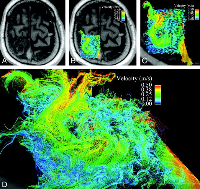
Disorganized, circulating blood flow within the AVM nidus. A is an axial magnitude section through the large left frontoparietal AVM. B has velocity data overlaid on this axial section. C is a magnified view of the AVM nidus with 3D velocity data from midsystole, as represented as a curved vector field, demonstrating somewhat disorganized, circulating flow. Note the high-velocity venous drainage in the superior sagittal sinus, which can be visualized through the semitransparent axial section. D is an oblique view at higher magnification.