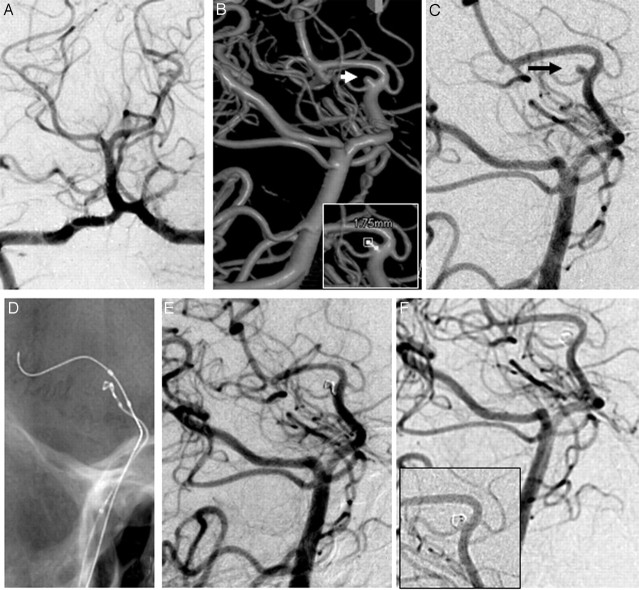
Fig 3.
A, DSA image (anteroposterior view). B, 3D image shows a small aneurysm at the origin of the posterior choroidal artery. C, DSA in the same angulation as the 3D image. D, Coil embolization with balloon assistance. E, Postembolization DSA. F, Follow-up DSA (note the coil artifact in the inset image).

