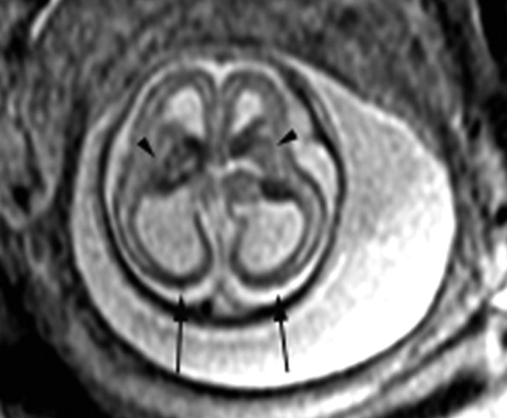
Fig 3.
Patient 3: 21-gestational-week fetus. Axial image demonstrates areas of hypointensity and hyperintensity involving the deep gray nuclei bilaterally (arrowheads), consistent with areas of hemorrhage and necrosis. There is bilateral ventriculomegaly with diffuse thinning of the parenchyma, with areas of hypointensity in the occipital and parietal lobes (arrows).

