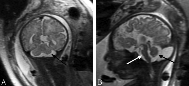
Fig 6.
Patient 28: 36-gestational-week fetus. A, Coronal image demonstrates a small left cerebellar hemisphere (arrow). The sulcation pattern is also diffusely abnormal with too-numerous infoldings of the cortex bilaterally. B, Sagittal image demonstrates a small pons (white arrow) and a small vermis (black arrow) as well as ACC.

