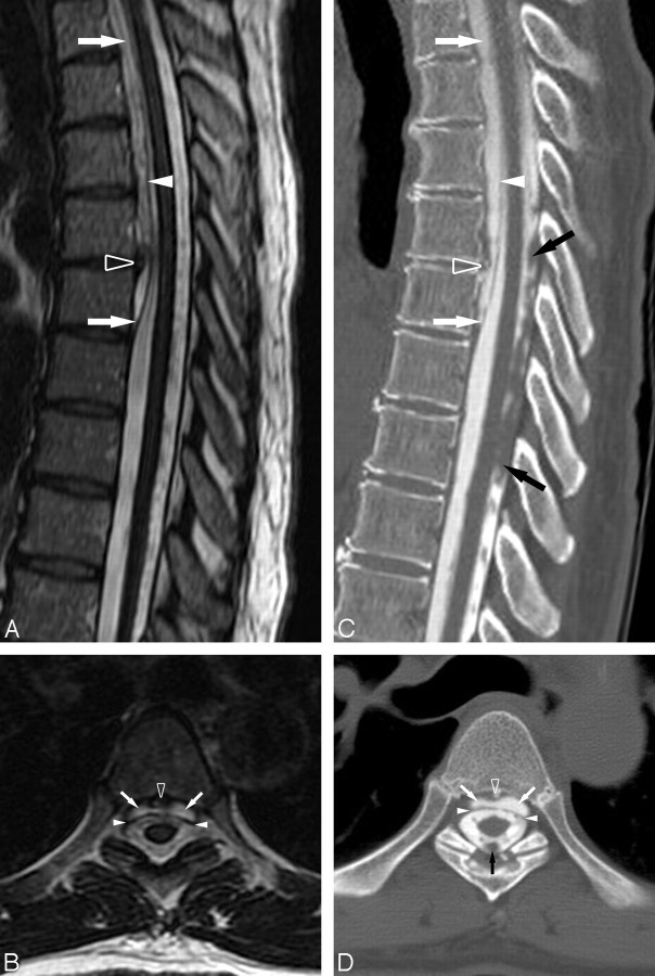
Fig 1.
A, Sagittal T2-weighted (TR, 3300 msec; TE, 100.8 msec) image of the thoracic spine defines the caudal aspect of the epidural fluid collection at T6–T7. A prominent central disk extrusion is present at T5–T6. B, Axial T2-weighted (TR, 2600 msec; TE, 103.1 msec) image through the T5–T6 disk extrusion further delineates the ventral epidural fluid collection displacing the dura posteriorly. Note the prominent T2 hypointensity on the cord surface secondary to superficial siderosis. C, D, Postmyelography CT images in similar planes and at analogous levels to the MR images demonstrate that the ventral epidural fluid collection is opacified by intrathecal contrast to a degree similar to CSF within the thecal sac, confirming the presence of an active CSF leak. The prominent T5–T6 disk extrusion is partially calcified. Ventral epidural fluid collection (white arrows), dura (white arrowheads), T5–T6 disk extrusion (open arrowhead), and subarachnoid clot (black arrows) are shown.

