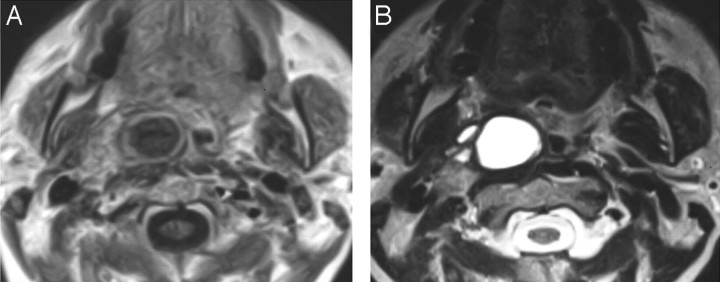
Fig 2.
A 52-year-old woman being evaluated with MR imaging for an unrelated indication. An incidental right parapharyngeal space mass was identified. A, Gadolinium-enhanced axial T1-weighted MR image shows a multiseptate cystic rim-enhancing mass in the right parapharyngeal space. B, Axial T2-weighted MR image shows the multiseptate parapharyngeal mass. The patient went directly to surgical resection, which identified metastatic papillary cancer in a high level II lymph node in the parapharyngeal space. Subsequent thyroidectomy identified a 4-mm papillary carcinoma in the thyroid gland. No other pathologic nodes in the neck were identified on MR imaging.

