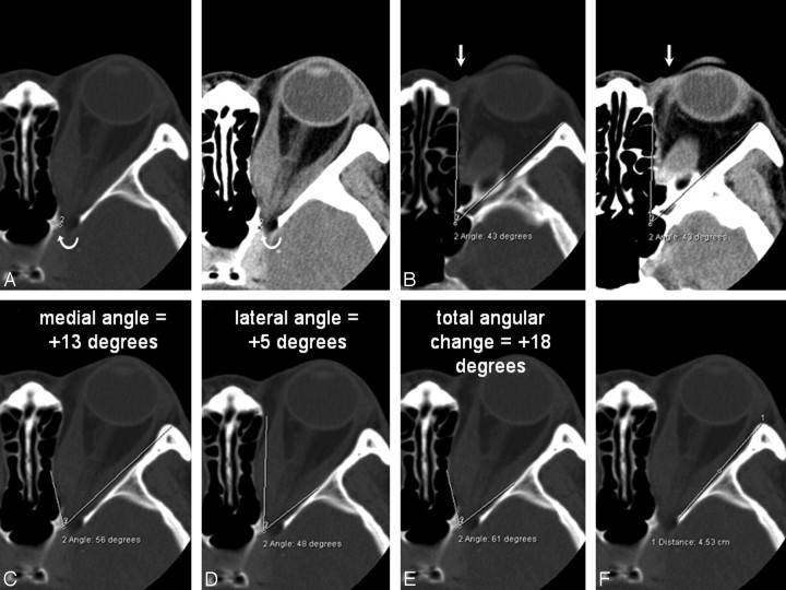
Fig 1.
A and B, Axial CT scans (bone window on left, soft tissue window on right) showing the orbital apex point indicated by the curved arrow (A) and the orbital rim angle (B). A, The apex point is defined as the anterolateral border of the groove in the sphenoid body formed by the intracavernous portion of the internal carotid artery (labeled 2) on the section just inferior to the anterior clinoid process. B, The orbital rim angle (43°) is measured at the level of the medial palpebral ligament (arrow). C−E, The same axial section containing the bulk of the medial and lateral rectus muscles shows the medial wall angle (C), the lateral wall angle (D), and the orbital apex angle (E). For the medial and lateral walls, the angle that best describes the widest bony point of the orbital wall around the muscular bellies is recorded. The angular change is recorded as positive if the resultant angle is wider than the orbital rim angle, and negative for narrower angles. F, The length of the lateral orbital wall is measured on the section just inferior to the anterior clinoid.

