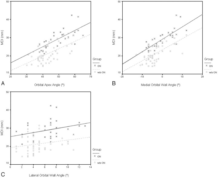
Fig 3.
Scatterplots of the MDI and orbital apex angle (A), MDI and medial wall angle (B), and MDI and lateral wall angle (C) in patients with and without ON. Greater muscular enlargement is accompanied by wider orbital angles with or without (w/o) ON. For identical MDI and orbital angles, the orbital angles are narrower and MDI greater in patients with ON, respectively. Within the borderline MDI range of 22–30 mm in B, 15 of 26 patients with zero or negative medial angles (57.7%) had ON (P = .07), compared with 1 of 6 patients with positive medial angles (16.7%).

