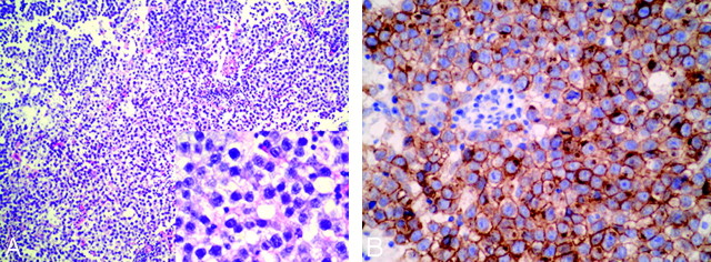
Fig 4.
Photomicrographs of histopathology of the right BG germinoma in case 4. A, Standard HE coloration (original magnification × 100; insert figure: original magnification ×400) shows that the tumor is composed of large cells with vacuolated cytoplasm and round or ovoid nuclei with prominent nucleoli. Lymphocytic element admixes with tumor cells focally. B, Immunolabeling of germ cells with CD117a (c-kit) is positive (original magnification ×400).

