
Fig 4.
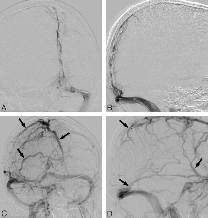
Frontal oblique and lateral angiographic views (A,B) demonstrate extensive SSS, torcular, and TS filling defects in keeping with DVST. After balloon thrombectomy and 7 days of intrasinus heparin infusion, despite only minimal recanalization of the SSS, robust cortical venous flow (arrows; C,D) is evident in this neurologically intact patient. Note recanalization of the TS and SS.