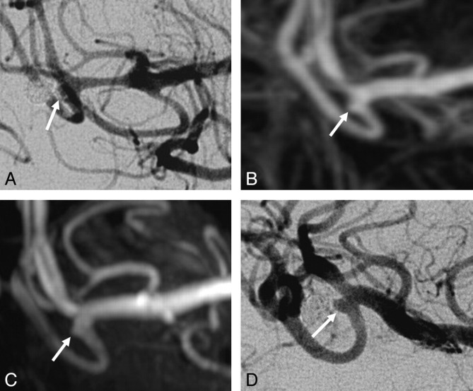
Fig 3.
Disagreement between both TOF-MRA and CE-MRA with angiography on the occlusion of a middle cerebral artery aneurysm. A, Angiogram obtained immediately after coiling shows a small neck remnant (arrow). B, Follow-up TOF-MRA at 6 months shows a small neck remnant (arrow). C, Follow-up CE-MRA at 6 months shows a small neck remnant (arrow). D, Follow-up angiogram shows incomplete occlusion (arrow). Because the geometry of the reopened aneurysm was unfavorable, this patient was not retreated but was subjected to extended follow-up.

