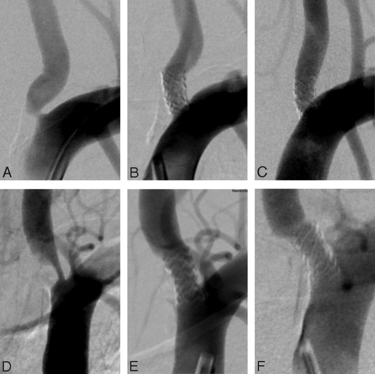
Fig 1.
Representative angiographic images from patients with significant vertebral ostial stenosis. A–C, Preprocedural (A), postprocedural (B), and 5-month follow-up (C) images in a 62-year-old male patient. D–F, Preprocedural (D), postprocedural (E), and 4-month follow-up (F) images in a 68-year-old male patient with significant stenosis of the left vertebral ostium.

