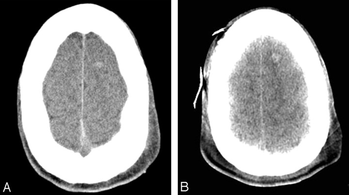
Fig 4.
Axial CT images at the centrum semiovale level show a small left frontal hemorrhage corresponding to shear injury. A, Image from a standard scanner. B, Image at a level comparable with that in A acquired on the portable scanner 8 hours later. Slightly better visualization of the hyperattenuated lesion with the portable scanner is likely due to interval clot retraction and development of perilesional edema.

