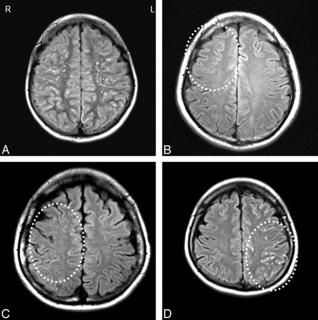
Fig 1.
The proliferative differences of ivy signs on FLAIR images between both hemispheres in each patient was rated as minimal, moderate, or marked. A, Symmetric ivy distribution. We defined the hemispheres as ivy symmetric when a symmetric ivy distribution was observed in both hemispheres. B, Minimal ivy proliferation along the cortical sulci dominantly in the right frontal lobe (dotted circle). C, moderate ivy dominance in the right hemisphere (dotted circle). D, Marked ivy distribution in the left hemisphere (dotted circle).

