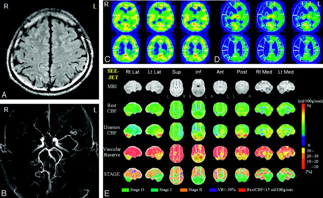
Fig 2.
Case of a 28-year-old man with unsymmetric ivy sign and decreased vascular reserve defined by quantitative SPECT analysis. He had sustained a left TIA. A, FLAIR image showed marked ivy sign in the right hemisphere. B, MRA stages in right and left hemispheres were III and II, respectively. C, Basal brain perfusion SPECT showed mildly decreased CBF in the right hemisphere. D, ACZ stress brain perfusion SPECT showed remarkably decreased CBF in the right hemisphere. E, Quantitative analysis of basal/ACZ stress brain perfusion SPECT. Lower 4 columns show 3D-SSP format view sets of rest CBF, Diamox CBF, vascular reserve, and staging by JFT study from top. Vascular reserve was impaired in most of the right ACA and MCA and in part of the left ACA and MCA territories. The proportions of the stage II area in the right and left hemispheres were 64.9% and 57.9%, respectively. Vascular reserve means in the right and left hemispheres were −9.63 and 2.80, respectively. Vascular reserve less than −30% areas were seen scattered in the right hemisphere. The patient is free from symptoms after cerebral revascularization on the right side.

